Why Composite Bonding Fails | 8 Causes | ATBIO
Why composite bonding fails is rarely explained by one material property alone. Direct composite restorations operate as a system: diagnosis, tooth substrate, isolation, surface treatment, adhesive, composite placement, light curing, finishing, occlusion, and patient maintenance all contribute to the result.
Clinical reviews identify fracture, aesthetic deterioration, caries, and retention problems among the reported reasons for intervention in anterior composite restorations. The relative importance of each factor varies by restoration type and patient. This guide organizes eight common failure pathways into practical checks, while distinguishing what may be repaired from what requires broader reassessment.
The recommendations below are general. Clinicians should follow the current instructions for each etchant, adhesive, composite, repair system, and curing device.
01 — Cause 1: Incomplete Diagnosis or Unsuitable Case Selection
A technically sound restoration can still have a poor prognosis if the underlying problem was not correctly defined. Active caries, inadequate enamel support, a large functional defect, severe parafunction, periodontal inflammation, an unfavourable margin location, or an aesthetic expectation that direct composite cannot predictably meet can all be mislabelled later as “bonding failure.”
Prevention: assess caries activity, remaining tooth structure, enamel availability, occlusion, guidance, wear pattern, periodontal condition, shade, and patient expectations before treatment. Document limitations and consider whether direct composite, an indirect restoration, orthodontic movement, bleaching, no treatment, or a staged approach better fits the case.
Repair decision: do not repair repeatedly until the original diagnosis and failure mechanism have been reviewed. A localized defect may be repairable, but disease, structural overload, or an unsuitable design must be managed first.
02 — Cause 2: Saliva, Blood, or Moisture Contamination
Adhesive bonding depends on controlled interaction between the tooth surface and the adhesive system. Saliva, blood, crevicular fluid, hemostatic agents, or other contaminants can reduce dentin bond strength, with the effect depending on when contamination occurs and which adhesive is used. The defect may not be visible when the restoration is finished.
Prevention: use rubber dam isolation when feasible or a well-planned alternative with retraction and suction. Prepare the field before etching so isolation is not improvised during the most technique-sensitive step. If contamination occurs, identify the stage and follow the manufacturer's validated cleaning and reapplication protocol. Water rinsing alone is not an adequate universal response for every material and stage.
Repair decision: a completely detached restoration, a localized marginal defect, and contamination discovered during placement are different situations. Rebonding or repair requires fresh substrate assessment and a documented surface-treatment sequence; simply placing adhesive over an unknown contaminated interface is not a predictable repair.
03 — Cause 3: Incorrect Etching or Substrate Management
Enamel and dentin are not interchangeable bonding substrates. Etching that is incomplete on enamel can limit micromechanical retention, while inappropriate dentin treatment can create sensitivity or a poorly infiltrated interface. Sclerotic dentin, uncut enamel, exposed cementum, existing composite, and ceramic each require their own surface strategy.
Prevention: identify every substrate at the margin and use the etching mode specified for the selected adhesive. Control contact time, rinsing, dentin moisture, agitation, evaporation, and application thickness according to the instructions. A thixotropic etchant gel can support precise placement, but product handling and clinical control remain essential.
Repair decision: sensitivity or marginal staining should be diagnosed before treatment. Surface polishing may be enough for a superficial issue; a defective adhesive interface, caries, or unsupported structure may require partial or complete removal.
04 — Cause 4: Adhesive Application Errors or Incompatibility
Modern adhesives can simplify inventory, but they are still technique dependent. Insufficient active application, incomplete solvent evaporation, pooling, over-thinning, inadequate curing, or combining materials without documented compatibility can reduce performance. “Universal” describes application flexibility; it does not mean that any sequence or material combination is validated.
Prevention: standardize one documented protocol, train the team on the exact sequence, protect bottles from contamination and inappropriate light exposure, observe storage and expiry requirements, and verify compatibility when a chemical-cure or dual-cure material is part of the workflow. Apply and air-thin for the specified times instead of relying on visual appearance alone.
Repair decision: if a localized area is clinically suitable for repair, prepare the aged composite and exposed tooth substrate separately. Roughening, cleaning, silane or primer, adhesive, and fresh composite may be required in different combinations depending on the surface and repair system.
05 — Cause 5: Inadequate Light Curing
Resin materials reach their intended properties only when the photoinitiator receives sufficient compatible radiant energy. Output at the light tip, wavelength, exposure time, distance, angulation, increment thickness, shade, opacity, and access geometry all influence polymerization. A device can illuminate the surface yet deliver inadequate energy to the deepest or most distant area.
Prevention: keep the light guide clean, verify device output with an appropriate maintenance program, position the tip close and perpendicular where possible, stabilize it during exposure, and cure from additional directions when indicated. Follow the material's stated increment and exposure requirements; do not assume that a higher advertised irradiance automatically compensates for distance, angulation, or incompatible wavelength.
Repair decision: a restoration suspected of inadequate deep cure cannot be predictably corrected by adding extra light days or weeks later. Clinically soft, unstable, or extensively defective material generally requires removal and replacement after diagnosis. A discrete surface or margin defect may follow a different repair pathway.
06 — Cause 6: Poor Increment Control, Anatomy, or Finishing
Exceeding the material's increment limit can compromise curing and increase the risk of voids or poorly controlled shrinkage stress. Inadequate adaptation, overbuilt contacts, thin unsupported edges, rough margins, and incomplete polishing introduce additional biological, mechanical, and aesthetic problems.
Prevention: place increments within the documented thickness, adapt each layer without trapping voids, design the restoration for the defect and load, and reproduce cleansable contours and contacts. Finish margins flush, preserve anatomy, and polish with a complete sequence. For larger posterior cases, the posterior composite restoration workflow provides a broader placement framework.
Repair decision: small voids, chips, or rough margins may be refinished or repaired if the remaining restoration is sound. Generalized under-curing, extensive open margins, caries, or an unstable restoration usually requires a more comprehensive intervention.
07 — Cause 7: Occlusal Overload and Parafunction
Fracture is a frequently reported reason for failure in anterior composite studies. Restoration size, tooth position, available enamel, incisal design, heavy contacts, deep overbite, edge-to-edge function, bruxism, and habits such as nail biting all change the load placed on a bonded restoration.
Prevention: evaluate static and excursive contacts before treatment and again after finishing. Design transitions and material bulk to avoid thin, unsupported composite where possible. Discuss behaviour modification and prescribe an occlusal appliance when clinically indicated. Material selection matters, but no composite or adhesive eliminates the effects of an uncontrolled load.
Repair decision: a localized chip is often a candidate for conservative repair when the remaining restoration is sound. Correct the contact, design, or habit that contributed to fracture before rebuilding the missing area; otherwise, repeat failure remains likely.
08 — Cause 8: Surface Aging, Caries Risk, and Aftercare
Loss of gloss, surface staining, marginal discoloration, plaque retention, recurrent caries, and gradual wear can develop over time. These findings do not all mean the same thing. A superficial stain may be removable by polishing, while a progressive gap or carious lesion requires diagnostic and restorative management.
Prevention: provide clear instructions on oral hygiene, interdental cleaning, staining exposure, hard-object habits, and recall. Use non-abrasive home-care advice appropriate to the patient. Review margins, surface texture, caries activity, and occlusion periodically. The earlier composite bonding longevity guide explains the maintenance factors in more detail.
Repair decision: distinguish refurbishing, resealing, repair, and full replacement. Evidence comparing repair with replacement is limited in certainty, but repair can preserve sound tooth structure when the defect is localized and the remaining restoration is acceptable.
09 — Composite Bonding Failure Checklist
Before treatment: diagnosis, caries and periodontal control, substrate, remaining tissue, occlusion, habits, shade, and expectations.
During isolation: rubber dam or equivalent control, retraction, clean field, and a defined response to contamination.
During adhesion: correct etching mode, active application, solvent evaporation, compatible materials, and full adhesive cure.
During composite placement: appropriate viscosity, controlled increments, adaptation, anatomy, and adequate material bulk.
During curing: clean tip, compatible wavelength, verified output, close orientation, stable exposure, and all required directions.
At finishing: smooth margins, cleansable contacts, polished surface, and verified static and excursive occlusion.
At recall: disease activity, symptoms, stains versus gaps, chips, surface wear, patient habits, and maintenance needs.
Consistency makes failures easier to investigate. A clinic that records materials, lot numbers, curing device, exposure sequence, isolation, and occlusal findings can identify patterns that would remain invisible when cases are documented only as “bonding completed.” The foundational sequence is reviewed in the complete composite bonding guide.
10 — Frequently Asked Questions About Composite Bonding Failure
1. Why does composite bonding chip?
Chipping can relate to occlusal overload, parafunction, unsupported design, inadequate material bulk, trauma, incomplete polymerization, or a combination of factors. The fracture pattern and remaining restoration should be assessed before deciding on repair.
2. Does marginal staining mean the bond has failed?
No. Superficial stain and roughness may be managed by professional finishing and polishing. A progressive gap, recurrent caries, sensitivity, floss catching, or loss of retention requires further diagnosis. Color alone cannot establish interface failure.
3. Can a failed composite restoration be repaired?
Many localized defects can be repaired when the remaining restoration and tooth are sound and the cause is manageable. Repair suitability depends on defect size, disease, substrate, access, restoration history, and patient risk. Extensive defects or caries may require replacement.
4. Can a weakly cured restoration be cured again later?
Additional immediate curing may be appropriate when an exposure was interrupted and the material protocol allows it. A restoration that is clinically under-cured throughout its body after service cannot be assumed to recover predictably from later surface exposure and may require replacement.
5. Is rubber dam mandatory for every composite bonding case?
Rubber dam is a highly effective isolation method, but case design and clinical circumstances vary. Whatever method is selected must provide reliable contamination control and access throughout the adhesive sequence.
6. Does choosing a stronger composite prevent every fracture?
No. Material properties are only one part of performance. Diagnosis, preparation design, adhesive protocol, curing, restoration geometry, occlusion, parafunction, and maintenance remain important.
11 — References
Demarco FF et al. Anterior composite restorations: A systematic review on long-term survival and reasons for failure. https://pubmed.ncbi.nlm.nih.gov/26303655/
Shah YR et al. Long-term survival and reasons for failure in direct anterior composite restorations: A systematic review. https://pmc.ncbi.nlm.nih.gov/articles/PMC8989165/
Mendes LT et al. Risk of failure of repaired versus replaced defective direct restorations in permanent teeth: a systematic review and meta-analysis. https://pubmed.ncbi.nlm.nih.gov/35362754/
Bourgi R et al. Effect of contamination and decontamination methods on the bond strength of adhesive systems to dentin: A systematic review. https://pubmed.ncbi.nlm.nih.gov/37395344/
Corrêa A et al. Polywave LEDs increase the degree of conversion of composite resins, but not adhesive systems: a systematic review and meta-analysis of in vitro studies. https://pubmed.ncbi.nlm.nih.gov/39982569/
12 — About ATBIO
This article is published by ATBIO, the dental-materials brand of AT&M Biomaterials Co., Ltd.
ATBIO (AT&M Biomaterials Co., Ltd.) manufactures composite resins, dental adhesives, etchant gels, cements, impression materials, endodontic products and related consumables, with OEM and private-label support for distributors. Request current product-specific instructions, conformity documents and quality-system certificates, and verify their scope for the intended product and destination. Where CE marking is applicable, its scope must be checked for the specific device. ISO 13485 concerns quality management; it does not establish clinical efficacy or authorization to sell in every market.
This article provides general educational information and does not replace diagnosis, individualized treatment planning, professional clinical judgment, or the instructions for use of a specific product. This page does not claim unconditional sale authorization in every country.
13 — Related Products
NANOFIL® Bonding Agent — a light-cured dental adhesive for professional evaluation according to its current instructions.
NANOFIL® Etchant Gel — a phosphoric-acid etchant for compatible enamel and dentin conditioning protocols.
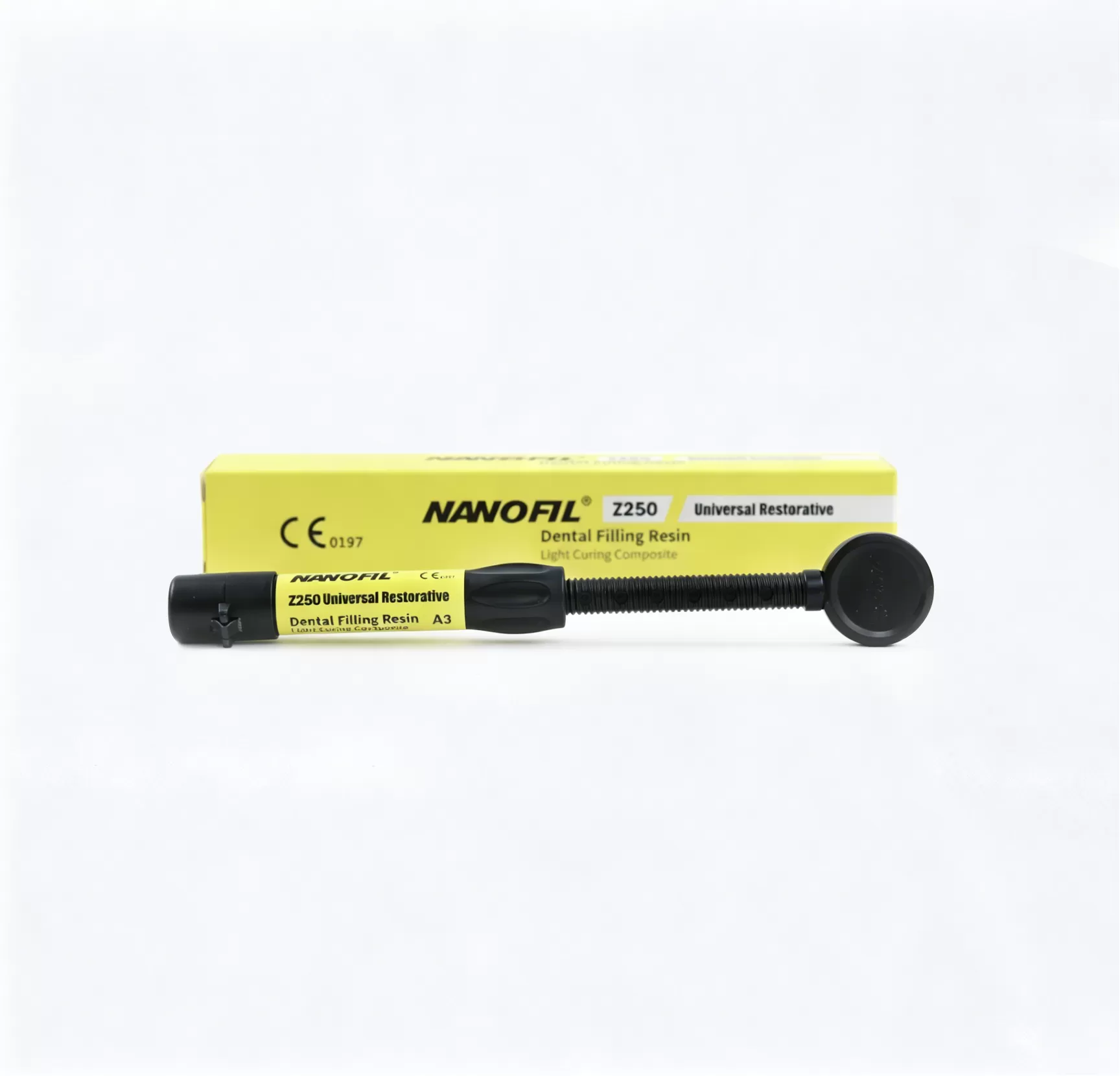
NANOFIL® Z250 Universal Restorative Composite — a universal restorative composite for use within its documented indications and curing protocol.
Build a repeatable bonding workflow around compatible, documented materials. Request a quote or contact ATBIO for current instructions, technical documents, samples, and distributor support.
Do you prefer online learning?
Live webinars and professional tips — learn when, where and how.
Access our online learning opportunities